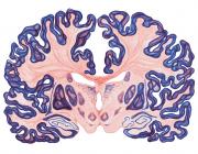
Retinotopic organization of human ventral visual cortex.
Publication Year
2009
Type
Journal Article
Abstract
Functional magnetic resonance imaging studies have shown that human ventral visual cortex anterior to human visual area V4 contains two visual field maps, VO-1 and VO-2, that together form the ventral occipital (VO) cluster (Brewer et al., 2005). This cluster is characterized by common functional response properties and responds preferentially to color and object stimuli. Here, we confirm the topographic and functional characteristics of the VO cluster and describe two new visual field maps that are located anterior to VO-2 extending across the collateral sulcus into the posterior parahippocampal cortex (PHC). We refer to these visual field maps as parahippocampal areas PHC-1 and PHC-2. Each PHC map contains a topographic representation of contralateral visual space. The polar angle representation in PHC-1 extends from regions near the lower vertical meridian (that is the shared border with VO-2) to those close to the upper vertical meridian (that is the shared border with PHC-2). The polar angle representation in PHC-2 is a mirror reversal of the PHC-1 representation. PHC-1 and PHC-2 share a foveal representation and show a strong bias toward representations of peripheral eccentricities. Both the foveal and peripheral representations of PHC-1 and PHC-2 respond more strongly to scenes than to objects or faces, with greater scene preference in PHC-2 than PHC-1. Importantly, both areas heavily overlap with the functionally defined parahippocampal place area. Our results suggest that ventral visual cortex can be subdivided on the basis of topographic criteria into a greater number of discrete maps than previously thought.
Keywords
Journal
J Neurosci
Volume
29
Pages
10638-52
Date Published
08/2009
ISSN Number
1529-2401
Alternate Journal
J. Neurosci.
PMID
19710316

